Plant disease refers to a series of morphological, physiological and biochemical pathological changes in plants under the influence of biological or abiotic factors, which hinders the process of normal growth and development. Under the influence of pathogens, the physiological functions of the plants are dysregulated and the tissue structure is destroyed. Plant diseases are antagonistic symbiosis of host plants and pathogens, and their occurrence and prevalence are the result of the interaction between host plants and pathogens. Plant diseases mostly occur in the leaves of plants, thus affecting the photosynthesis of plant leaves. Beijing Ecotech Ecological Technology Co., Ltd. provides a comprehensive technical program for plant disease research, detection and evaluation for researchers in plant protection and plant diseases.
This technical solution mainly includes RGB imaging analysis, infrared thermal imaging analysis, and FluorCam multispectral fluorescence imaging analysis technology. RGB imaging analysis mainly performs morphological measurement and color grading analysis of plant leaves or fruits during development in the visible light band of 400-700nm; infrared thermal imaging analyzes the dynamic distribution of plant temperature to reveal the process of plant diseases and the dynamics of stomatal conductance FluorCam chlorophyll fluorescence imaging is a sensitive probe of plant stress, which can fully reflect the infection process of plant diseases, stress physiology and resistance.
RGB imaging analysis:
RGB imaging analysis technology is based on visible light plant reflection spectrum imaging to analyze plant morphology and color data, and can obtain the area, length, width, color grading, greenness index, etc. of plant leaves and lesions, and can be divided according to different colors, and Calculate the area and ratio of each area, etc., widely used in plant pathology research.
Infrared thermal imaging analysis:
Infrared thermal imaging analysis can measure the temperature of each pixel point online and perform various point, line, and area measurement displays. It is widely used in plant science (such as plant stomatal conductance analysis and research, plant stress, etc.), animal science (such as animal energy Metabolic analysis research, animal surface heat distribution measurement, etc.), agriculture and forestry science, ecology, industrial quality control and UAV remote sensing, etc.
FluorCam multispectral fluorescence imaging analysis:
FluorCam Multispectral Fluorescence Imaging System is an extended version of FluorCam Chlorophyll Fluorescence Imaging System. It can be used not only for dynamic imaging analysis of chlorophyll fluorescence, but also for long-wavelength ultraviolet ultraviolet light (320-400nm) to stimulate multispectral fluorescence of plant leaves (UV-MCF) Perform imaging analysis.
Applications:
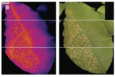
An article published in "Frontiers in Plant Science" by the Spanish Plant Biocytology and Biochemistry and Molecular Biology Research Center in February 2018, using RGB imaging, infrared thermal imaging and FluorCam multispectral fluorescence imaging on melon leaves infected with Dickeya dadantii Perform observation, detection and analysis.
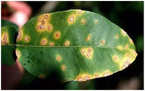
Worldwide, the main culprit causing major economic losses to crop production is the pathogenic bacteria D. dadantii. In the melon leaves, D. dadantii produces multiple spots of necrosis, necrosis of the entire infected area and chlorosis of surrounding tissues. The degree of these symptoms is gradually increasing from the day the disease appears. Several imaging techniques (chlorophyll fluorescence, multispectral fluorescence, and thermal imaging) provide information on the spatial and temporal changes of stomatal activity and primary and secondary metabolism of infected leaves. Use the data images provided by these imaging techniques to detect diseased leaves. It is a reliable method to detect the disease of the entire leaf through the model established on the leaf infection area. This method is especially suitable for the application of plant phenotype or precision agriculture.
Inoculate Dickeya dadantii bacteria on the second leaf of a three-week-old plant, inject a low-dose (LD) or high-dose (HD) bacterial suspension into the back of the leaf with a 1 ml syringe, and infect 4 or 6 areas, each The infected area is 1 cm2, and the infected area accounts for 5% of the total leaf area. For image analysis, each leaf defines three areas of interest: the infected area (I), the adjacent area (N), and the area farther away from the area I (D). Imaging measurements were performed 3 days and 7 days (dpi) after inoculation.
As shown in Figure 1, the change in symptoms caused by LD or HD after inoculation of melon leaves. Control and bacterial infection areas are displayed on RGB and leaf temperature images, as well as images calculated for Fv / Fm and F440 / F520 ratios. It can be seen from the figure that the two infections become more severe as the number of days increases; for the same number of days, HD infection is more severe than LD.
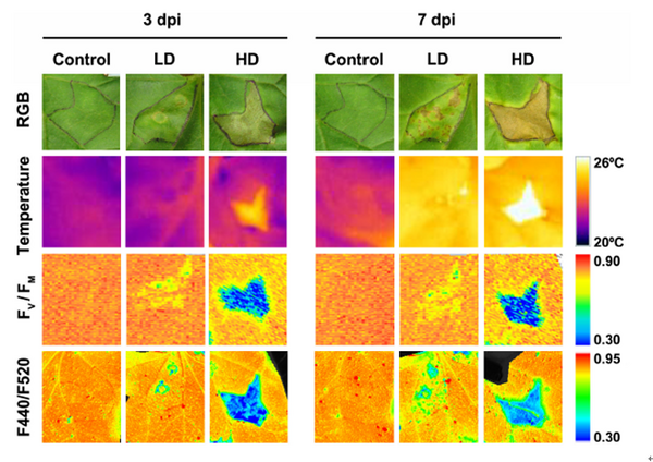
Fig.1 Disease changes of leaves after infection with D. dadantii
Leaf temperature is affected by D. dadantii to varying degrees. According to the different bacterial doses, among the 3 dpi LD infected leaves, only the temperature displayed in zone I was higher than the corresponding control; however, at 7 dpi, the entire leaf temperature was higher than that of the control group. In the case of HD infection, the average temperature of the infected leaves is higher than the average temperature of the control leaves. This effect is more severe at 7 dpi.
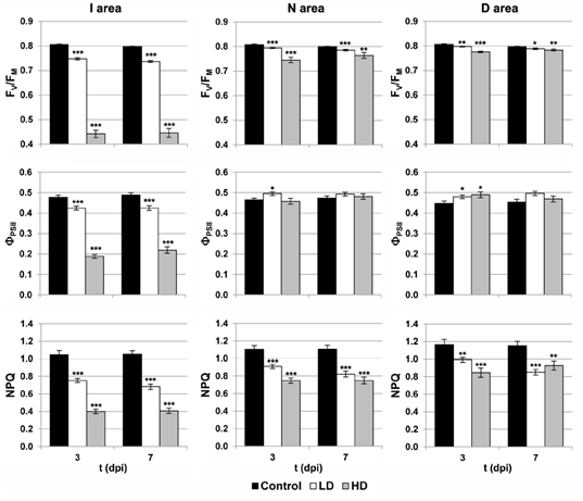
Figure 2 Chlorophyll fluorescence parameters of I, N and D regions of each leaf
Photosynthesis was affected by D. dadantii infection, as shown by the chlorophyll fluorescence kinetic parameters (Figure 2). The values ​​of Fv / Fm and NPQ were significantly reduced relative to the corresponding areas of the control leaves. This effect is found in each of the three areas analyzed by dpi and is more severe when HD is infected.
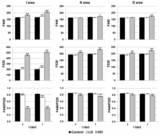
Figure 3 Multi-spectral fluorescence parameters of the I, N and D regions of each blade
MCF analyzed the activity of secondary metabolism (Figure 3). In the case of HD inoculation, the entire leaf showed significant changes at 3 and 7 dpi. In contrast, F440 showed that, compared to the control, the I area of ​​3dpi and the remaining area of ​​the 7dpi blades increased moderately. In the case of LD infection, F440 (7 dpi) and F520 (3 and 7 dpi) showed a small but statistically significant increase, but only in the I area. In addition, F440 / F520 only decreased significantly when infected.
The company is a company that mainly produces all kinds of luggage products, mainly handbag packaging, gifts, non-woven products, computer bags, travel bags, backpacks, cosmetic bags, Essential Oil Bags, camera bags, handbags, shopping bags, tool bags, fishing bags, business backpacks, shoulder bags, waist bags, student backpacks, trolley suitcases and other products; the company's business purpose: product specialization, price maximization, high-quality service; suitable for the needs of the market, in line with the needs of customers; the principle of reputable reputation, building image, commitment is gold, and winning the praise and recognition of the majority of consumers.
11 Sets Baby Care Products,New Baby Healthcare Kit,Baby Health Safety Kit,Newborn Baby Care Kit
Dongguan Junkai Packaging Products Co., Ltd , https://www.diystoragecases.com